
This was the Mother of all Headaches
Joe’s headache had lasted for days. He went to sleep with a headache, and he woke up with one

CT scan showing a brain aneurysm.
He said he couldn’t see clearly at times, felt tired all the time, and sometimes became confused about where he was. He was out shopping with his wife when he suddenly collapsed unconscious. Someone called the ambulance, and he was rushed into hospital.
Joe’s wife explained to the emergency team how he had been feeling and behaving. They suspected Joe had an aneurysm in his brain and had suffered a bleed. A CT scan quickly revealed the diagnosis. This was followed up with an angiogram. A catheter was inserted into Joe’s groin and using x-rays it was guided through his blood vessels into his neck, where the blood vessels supply his brain. A dye was injected, and x-rays taken. The image showed the aneurysm clearly.
The neurosurgeon told Joe and his wife that although the aneurysm hadn’t ruptured and was bleeding into his brain, he needed an operation quickly. Joe’s operation was scheduled for the next morning.
The neurosurgeon explained that Joe’s aneurysm is a weak or thin spot on an artery in the brain that balloons or bulges out and fills with blood
The bulging aneurysm can put pressure on the nerves or brain tissue. There is a risk that the aneurysm could rupture causing blood to flow into the brain tissue and this could cause a stroke, brain damage, coma, and even death. The surgeon explained that the operation was necessary but there were risks associated with it. These included:
Behaviour changes due to neurological injury
Blood clots
Brain swelling
Confusion
Infection
Seizures
Speech and vision problems
Stroke
General weakness
The surgeon explained that he would clip Joe’s aneurysm and he would perform a craniotomy
A small part of Joe’s skull would be removed. The dura, which is the outer covering of the brain is opened to expose the brain. Then using microsurgery, the surgeon would access the aneurysm through the natural spaces or anatomical divisions (‘fissures’) between the parts of the brain (‘lobes’) to try to avoid damage to brain tissue. The aneurysm is then carefully separated from the normal blood vessels and the brain, so can be treated. The surgeon explained that he would then attach A small metal clip, made from titanium to the neck of the aneurysm. This would seal the aneurysm from the blood supply so it couldn’t get any larger and rupture. He would then close the skull using small screws. The procedure would take anywhere between three to five hours.
The surgeon had explained the risks associated with the procedure, particularly the risk of postoperative neurologic deficit (PND). Examples of PND include motor weakness, sensory deficits, language difficulty, visual deficits, and confusion to name a few. If you look at the diagram below of the cortex of the brain, you can see which areas of the brain control the various motor and sensory functions. It’s easy to imagine how an aneurysm and subsequent surgery can lead to PND.
The motor and sensory areas of the cerebral cortex

The surgeon explained that to reduce the risk of PND they would use Intraoperative neurophysiological monitoring (IONM) during the operation

A typical IONM machine.
IONM protects patients by continuously monitoring the central nervous system (the brain, spinal cord, and nerves) when it is at risk during surgery. Electrodes are attached to the wrist, ankles, scalp, and sometimes to specific muscle groups. The electrodes record the response of the nervous system to electrical stimulus and can indicate changes in the functioning of the nervous system. If there are significant changes during the operation that could be due to the procedure, the surgeon is able to change the approach and avoid causing irreversible neurological damage.
Joe’s operation happened the next day and was a complete success with the aneurysm successfully clipped. Joe went home a few days after the operation. It took a few weeks for Joe to recover fully. He was told to expect to get headaches, maybe feel confused at times and get tired easily. He couldn’t drive for a few weeks, had to avoid exerting himself and was unable to play sport. At his six month follow up appointment Joe had fully recovered and could get on with his life.
Intraoperative neurophysiologic monitoring (IONM) is not just used for aneurysms
It is recommended for individuals at increased risk of neurological injury during surgical procedures such as spine and spinal cord surgery and brain and brain stem surgeries including craniotomy for tumour removal. One of the unfortunate complications of using IONM is that nerve stimulation can produce a powerful activation of the jaw muscles causing the patient to suffer tooth fracture and/or tongue and lip lacerations that sometimes require suturing.
As always in this area there is little published data on the incidence of bite injuries associated with this procedure. One study from 2012 reported an incidence of 0.63% with the majority being tongue injuries(1). A single hospital study from 2018 suggested that in clinical practice, bite injuries appear to occur more frequently than previously reported. This study reported an incidence of 6.5%(2). All studies and articles recommend the use of up to three soft bite blocks to protect the patient with frequent checks to ensure they remain in place. The blocks are placed on either side between the molars and a third is placed centrally.
Frequently bite blocks are made up in theatre before the surgery
Many practitioners use rolled pieces of gauze as bite blocks, but these have to be constructed, may be contaminated, may be difficult to insert, often are ineffective and can get caught on teeth and braces. Rolled gauze bite blocks also cost more than most people think. At Innovgas we have developed BiteMe which is a purpose designed, air-filled, soft plastic bite block. The combination of the soft plastic surrounding a closed air-filled space means that when a patient bites down, there are two forces opposing the bite making it an ideal bite block for this group of patients. Request a free sample through the simple form submission below and find out for yourself.
References:
The incidence of bite injuries associated with transcranial motor-evoked potential monitoring. Arvydas Tamkus, Kent Rice. Anaesth Analg 2012 Sep;115(3):663-7.
Bite injuries caused by transcranial electrical stimulation motor-evoked potentials' monitoring: incidence, associated factors, and clinical course. Sachiko Yata, Mitsuru Ida et al. J Anaesth. 2018 Dec;32(6):844-849.

Author: Niall Shannon, European Business Manager, Innovgas
This article is based on research and opinion available in the public domain.
Interested in a Free Sample?
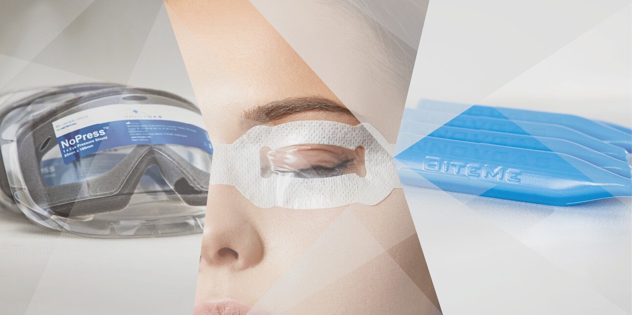
Free samples of NoPress, EyePro & BiteMe available upon request.
Conditions apply.